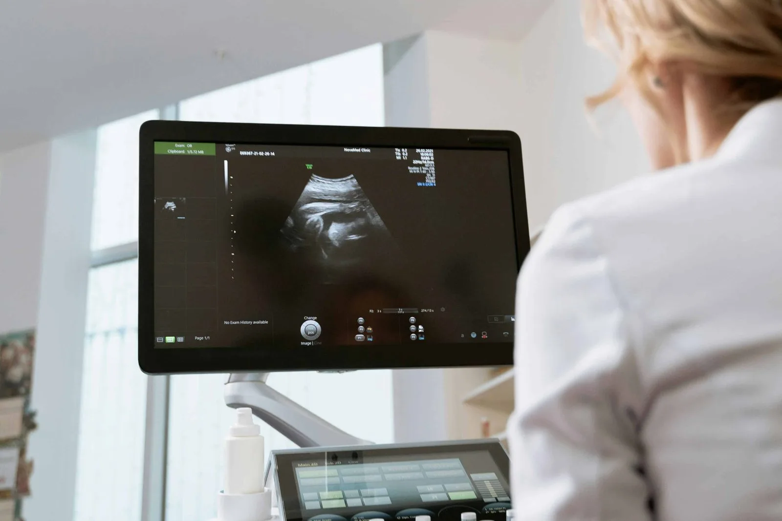
What a abdominal ultrasound shows
An abdominal ultrasound images the liver, gallbladder and bile ducts, pancreas, spleen, kidneys and the abdominal aorta. It is the first-line test for suspected gallstones, for abnormal liver blood tests, for kidney stones and cysts, and for following known findings over time. It uses sound waves, not radiation, and needs no contrast.
When it is ordered
- Pain in the upper abdomen or right side, or after meals.
- Abnormal liver enzymes or other liver blood tests.
- Suspected gallstones or gallbladder inflammation.
- Kidney stones, cysts, or abnormal kidney blood tests.
- Checking the size of the aorta.
- Follow-up of a known finding at the interval your provider sets.
What happens during the exam
You register at the Bells Corners desk about 10 minutes before your appointment. The sonographer confirms your identity and the requisition, explains the exam and asks you to uncover the abdomen; for some exams you change into a gown. Warm gel is applied and a hand-held transducer is moved over the area while images are recorded. You will be asked to take and hold deep breaths and to turn onto your side so each organ can be seen. The gel wipes off and you can eat as soon as you are done.
After the exam
The images are interpreted by a radiologist and the report is sent to your ordering provider, who reviews it with you. Allow several business days. If something needs prompt attention, the radiologist contacts your provider directly; the sonographer cannot discuss findings during the exam.
Bloodwork, X-ray or ECG on the same visit
Bells Corners is a full patient service centre: bring any lab requisition and have walk-in blood collection, X-ray or ECG done before or after your scan. Check our fasting guide if your bloodwork needs it; abdominal ultrasound and fasting bloodwork pair well on one morning visit.