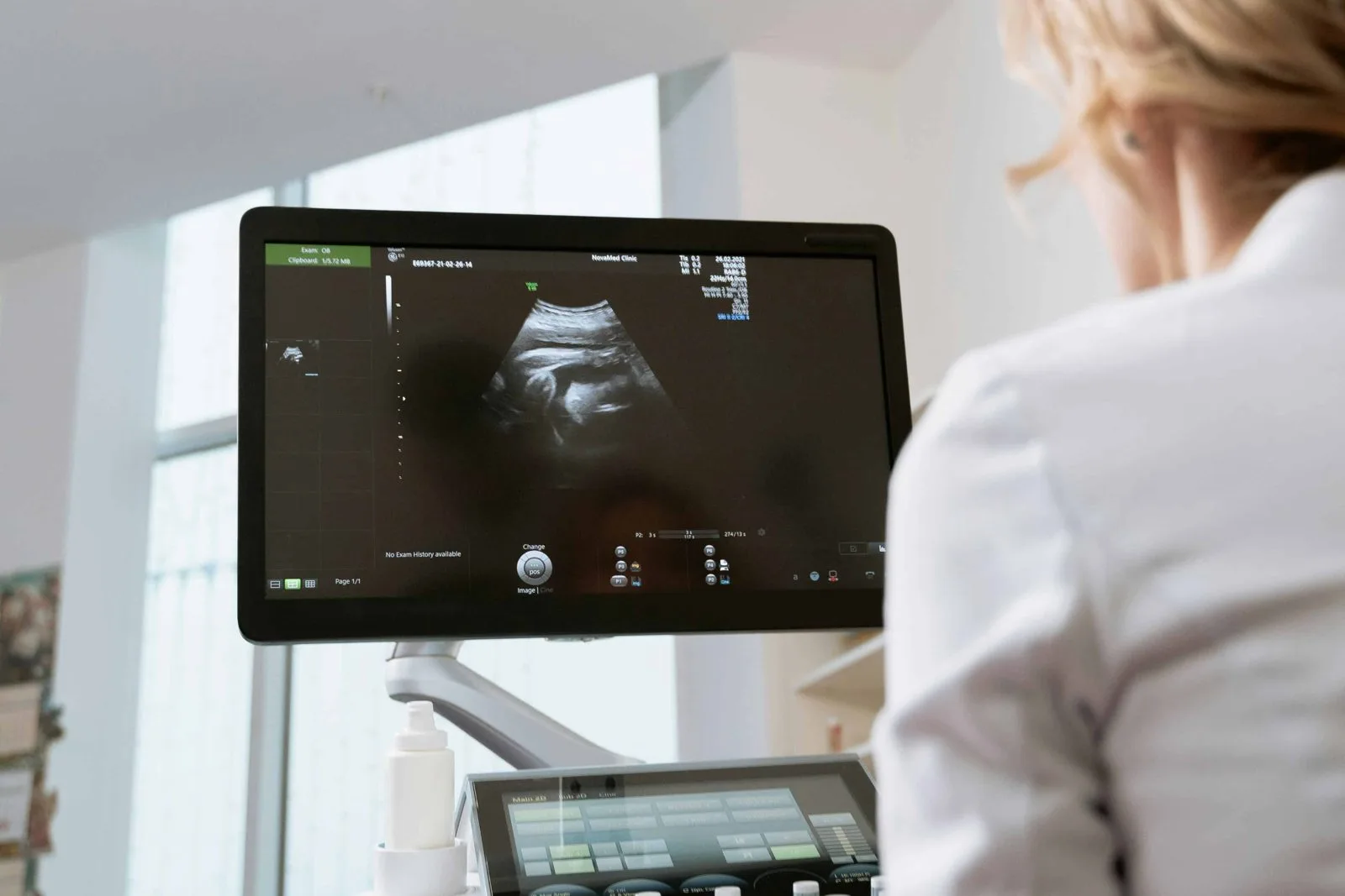
What a pelvic ultrasound shows
A pelvic ultrasound images the uterus, ovaries and bladder, or the prostate and bladder, through the lower abdomen. With a full bladder acting as a window, the sonographer measures the organs, looks for fibroids, cysts and other findings, and can check how completely the bladder empties. If your provider has requested a transvaginal view for more detail, the sonographer explains it before proceeding and you may decline.
When it is ordered
- Pelvic pain or pressure.
- Abnormal or heavy menstrual bleeding, or bleeding after menopause.
- Suspected fibroids or ovarian cysts, or to follow known ones.
- Urinary symptoms, or to measure how well the bladder empties.
- Prostate and bladder assessment in men.
- Follow-up of an earlier finding, at the interval your provider sets.
What happens during the exam
You register at the Bells Corners desk about 10 minutes before your appointment. The sonographer confirms your identity and the requisition, explains the exam and asks you to uncover the lower abdomen. Warm gel is applied and a hand-held transducer is moved over the area while images are recorded; you may be asked to hold your breath or change position. If a transvaginal view was requested, it is explained first and done only with your agreement, usually after you empty your bladder. The gel wipes off and you can return to normal activities immediately.
After the exam
The images are interpreted by a radiologist and the report is sent to your ordering provider, who reviews it with you. Allow several business days. If something needs prompt attention, the radiologist contacts your provider directly; the sonographer cannot discuss findings during the exam.
Bloodwork, X-ray or ECG on the same visit
Bells Corners is a full patient service centre: bring any lab requisition and have walk-in blood collection, X-ray or ECG done before or after your scan. Check our fasting guide if your bloodwork needs it; abdominal ultrasound and fasting bloodwork pair well on one morning visit.